At Evershine Optical, besides checking for the degree of your eyes, our eye examination includes the checking for eye diseases, especially when there is unexplained poor vision in one of both eyes. The most basic equipment use to do that is the slit lamp biomicroscope, which enables us to check the front part of the eyes as well as the back part of the eyes in a highly magnified view. We have also added the Icare Pro, which is used to measure the pressure of the eye to check for glaucoma as well as the Zeiss VIsucam 500 fundus camera which is helpful in visualizing the back part of the eye and for documenting and monitoring any changes.
Recently we have a 50+ year old customer who came in to make a pair of progressive lenses because his distance and near vision has deteriorated. He has perfect vision all his life and we presume because he didn’t need glasses, he never had an eye check before. During the eye examination, the vision in his left eye was found to be poorer than the right eye. He may not be aware of this because people seldom cover their eyes one at a time to check their own vision.
We check his eyes and did not see anything that could have caused the poorer vision in the left eye. So we decided to take a fundus photograph with the Zeiss Visucam 500 .
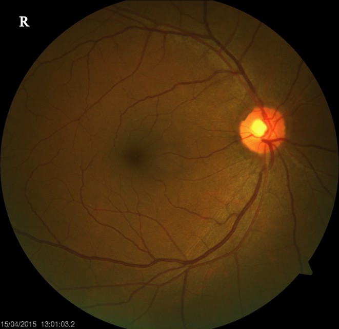
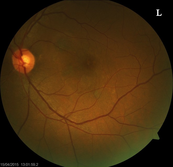
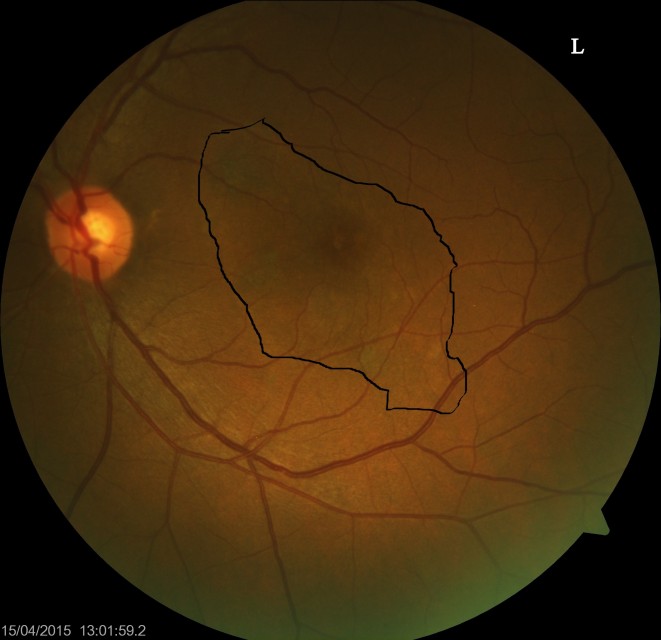
Although we are able to examine the fundus using a biomicroscope, a photograph allows us study it closely without much discomfort to the patient. With a casual glance, the right and left eye may look normal and healthy. However, when we study it closely, we notice that the left fundus is slightly different.
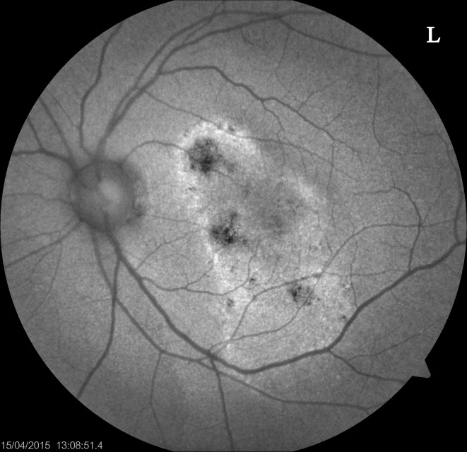
Examining the left photo, we can see a very faint demarcated area that is not seen the in the right eye. The Zeiss Visucam has a function to capture fundus autofluorescence (FAF) image of the retina. FAF imaging can provide information about the health and function of the retina.
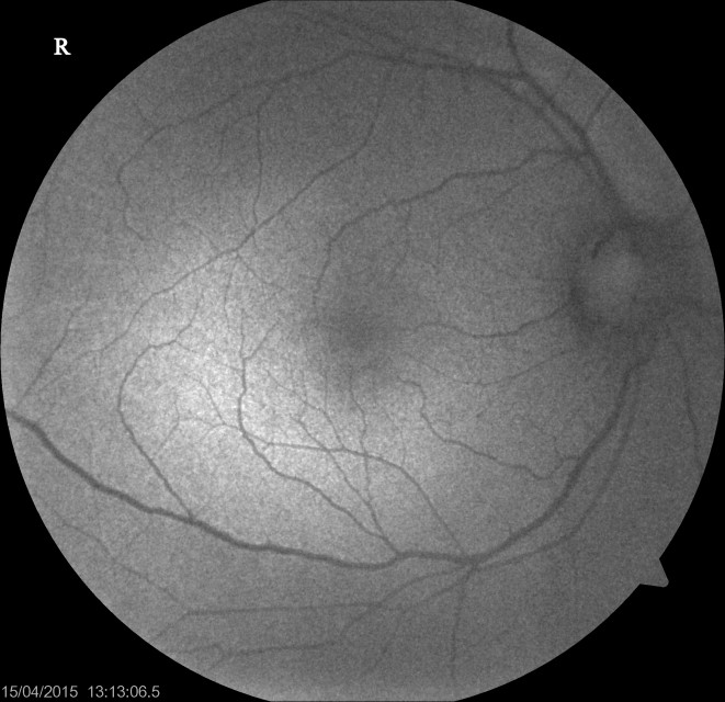
The FAF image shows changes in the retina that is not readily seen in a normal retinal photo. Below is a FAF image of the right eye
We believe the poorer vision in this patient’s left eye is due to the changes seen in the FAF image in the left eye. We are referring our patient to our trusted eye specialist partner for further examination and to see if there is any treatment to stop the progress of the disease.

